Copyright © 2012 All Rights Reserved to Gamma Knife Center
Nasser Institute
Tell me more about Gamma Knife Radiosurgery?
Gamma Knife is a method of treatment called radiosurgery.but what is radiosurgery?
Radiosurgery
Radiosurgery was invented by a Swedish Professor of Neurosurgery in 1951. It was he who first used the word radiosurgery. Dr. Leksell had a more than usually detailed education in how the nervous system worked and he thought that many operations did not show the brain the respect due to such a complicated and delicate tissue. So he dedicated his life to develop more delicate methods. The idea behind radiosurgery was that it was more elegant to perform an operation without opening the head. After much preliminary research Professor Leksell settled on Gamma Rays as the best instrument for performing this kind of surgery.
What are Gamma Rays
Two kinds of electromagnetic rays are commonly used in medicine. These are X-rays and Gamma Rays. The rays as rays are very similar. Where they differ is how they are made. X –rays are made in a man made machine and Gamma Rays are produced by the breakdown of a radioactive substance (or isotope). As Dr. Leskell’s machine uses Gamma Rays, it is a Gamma machine.
Why Gamma Knife
The definition Dr. Leksell gave to radiosurgery was a single session radiation treatment where radiation is delivered with surgical precision. Surgery is carried out with a knife. Since his ma-
chine uses Gamma rays it was a simple extra step to call it the Gamma Knife. As the reader can see this 19 ton machine does not look at all like a knife. But it is the machine’s function not its appearance, which justifies the use of the term.
What is it for
Today neurosurgery is very much safer than it was even 30 years ago. Better anaesthesia and the improved lighting and view of the operation gained by using an operating microscope have been major contributions to this increased safety. Even so the brain remains a major challenge for the surgeon. There are still areas where he does not want to go. The places inside the brain where there is most risk are the deep central portions. In addition the skull base or part of the skull the brain sits on is also a hazardous area.
Danger Areas
There are reasons that these two areas are particularly risky. The central part of the brain can be reached with accuracy today so it is not just the depth that is the problem. The difficulties are rather related to the way in which the brain is constructed. It is a reasonable analogy to suggest that the surface of the brain is like a computer while the deeper portions are like the wires. The computer may be more complex in its nature but its components are spread
out so that a small amount of damage may pass unnoticed. The central wiring is very compressed so that the wires (nerves) to many parts of the body are all compressed together in a tight space. This means that even a slight damage may have very marked effects on the patient. Thus the centre of the brain is a risky area for the surgeon. (See the diagrams below). The skull base is risky because it is hard to access parts of it. However, the major difficulties are related to the nerves and blood vessels, which enter and leave the skull through its base. Damage to these will give the patient long lasting significant losses of function.
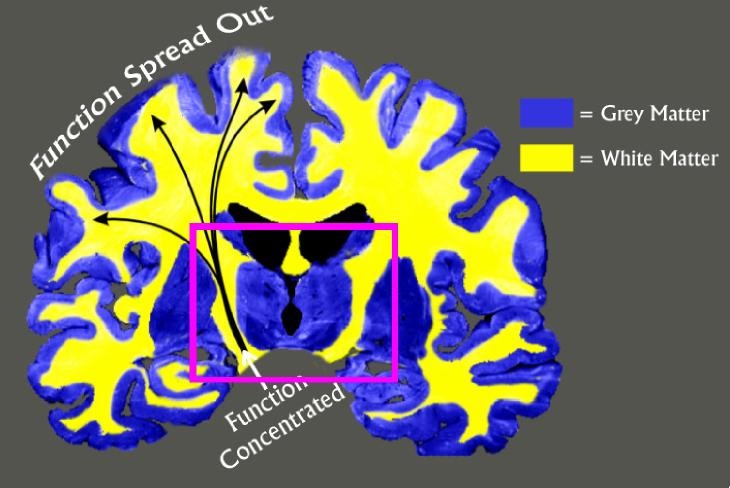
This diagram shows how the nerve fibres are arranged in the brain. The grey matter
(blue in this diagram) is like the brain’s computer. The white matter (yellow in
this diagram) is the brain’s wiring. As the diagram shows the wiring is spread out
on the surface but concentrated in the deep parts of the brain. Thus while the white
matter is not so complicated as the grey matter, damage in the central can affect
far more function than damage on the surface. This means the patient will suffer
far more paralysis. 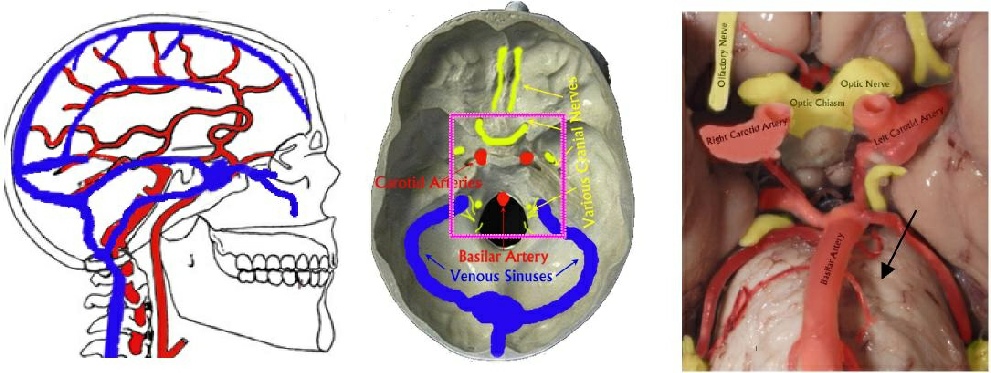
A B C
- Is a diagram of the arrangement of the arteries and veins in the head. It is clear that they enter and leave through the skull base.
- Is a diagram of the skull base showing where the red arteries blue veins and yellow nerves leave the skull. These are firmly since they pass through the bone and are often in the surgeon’s way. Damage to them can produce profound damage to the patient’s function. In this picture you are looking from above down
- This is an enhanced picture of the base of the brain. In this picture you are looking from below up. Arteries and nerves are shown. The brain stem contains the continuation of the fibres running to and from the brain in addition to collections of nerves for controlling the feeling and movement in the head. The picture shows how close everything is to everything else. Thus there is very little room for error. The tiny blood vessels visible branching from the basilar artery are each and every one of them vital. Damage to just one could give massive loss of function.
The dotted lilac rectangle again indicates the regions where the dangers of surgery are greatest. Thus this diagram illustrates why the skull base is also an area where open surgery can be more hazardous because of the way the body is made. The possibilities today are much greater than 20 years ago but there are still definite limitations to what may be achieved with safety. Thus, a most important role of Gamma Knife radiosurgery is to take over where the surgeon does not want his knife to go. The technique helps the surgeon and increases the chances of safe effective treatment
Today!
While modern skull base surgery is a well-developed and skilful technique there are still many diseases, which cannot be completely treated because of the risks.
The risks of operating in the centre of the brain and underneath it at the skull base are major reasons for the value of radiosurgery. This method avoids
manipulating sensitive central parts of the brain or blood vessels and nerves leaving it are under surface. Thus, radiosurgery permits safer surgery allowing the surgeon to stop operating before he gets into a really risky situation. Radiosurgery can treat the part of the tumour or other disease process, which remain after surgery.
What has it achieved?
The early days
In the first years Professor Leksell was very cautious and scientific in his approach. Only a few patients were treated as the method gradually proved itself in the treatment of living people.
The watershed.
After many years in Stockholm, a couple of units were placed in departments run by ex pupils of Leksell. One was in Buenos Aires and one in Sheffield. Then the Unit in Pittsburgh started operations in 1986 and the milieu changed quickly.
The Pittsburgh group treated many patients and reported results with great clarity and honesty. Anyone could relate to what they were doing. And to be honest the location of Pittsburgh in the USA was important in spreading the use of radiosurgery.
Now Routine
Today the situation is very different. It is almost impossible to hard to open a major professional journal on neurosurgery without finding an article devoted to some aspect of radiosurgery.
The nature of the method
How does the Gamma Knife work? It is surprising how many people still think that it is some kind of laser. While medically qualified readers need more detail and can find it in the specialist literature, the patient is not interested in such detail. Nonetheless the patient needs to know the principle.
It is useful to remind people of the way a magnifying glass focuses the sun’s rays. If people lie out in the sun they get tanned but they do not burst into flames. However, if the sun’s rays are focused it is easy to start a fire.
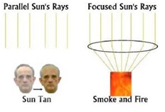
The Gamma Knife focuses radiation, which is powerful enough to kill living cells. This makes the effect much more powerful. At the same time the machine allows the doctor to sculpt the radiation field to match the shape of the target being treated. Thus, the essence of the method is very accurately applied focused radiation. How much damage the rays do is determined by how long the patient is kept at the radiation focus. This is just like the magnifying glass which can discolour a piece of paper or make it burst into flames all according to how long it is kept at the focus of the sun’s rays.
The safety of the method
To date it is believed that no-one has died from Gamma Knife radiosurgery. Some people have suffered from minor complications but these have decreased in number and severity with continuing improvements in imaging methods and treatment planning.
| Who we are |
| Gamma Knife Staff |
| Tell me more |
| FAQ |
| Photo Gallery |
| Video Gallery |